Patient with marked claustrophobia and left shoulder pain with limitation of movement.
A 51-year-old male patient presented with acute left shoulder pain with limited mobility for 2 weeks.
Referral for MRI was made with the suspicion of rotator cuff rupture. No previous operations in the left shoulder joint.
The patient suffers from pronounced claustrophobia. A conventional MRI in a “tube system” was not tolerated by the patient.
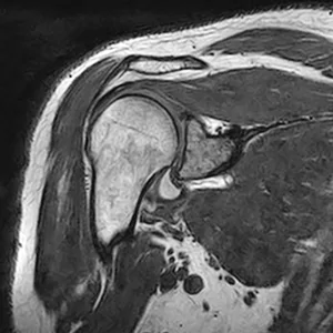
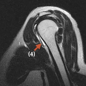
a) Coronary 3D sequence
b) Coronary T1-weighted
c) Coronary STIR sequence
d) Sagittal T2-weighted
e) Sagittal T2-weighted
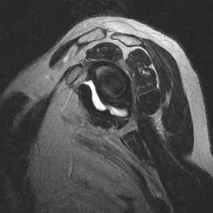
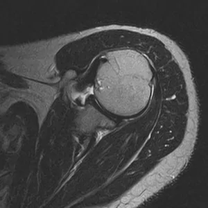
f) Transversal T2-weighted
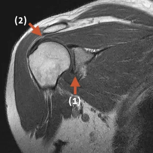
Overall, the shoulder joint is age-matched with incipient osteoarthritis and the onset of osteophytes (1) and joint space narrowing with a small joint effusion.
The AC joint shows non-activated osteoarthritis.
The subarachnoid space (2) is moderately constricted with a width of 6 mm, and the supraspinatus tendon is signal-altered in the muscle-tendon junction (3), but without interruption of continuity.
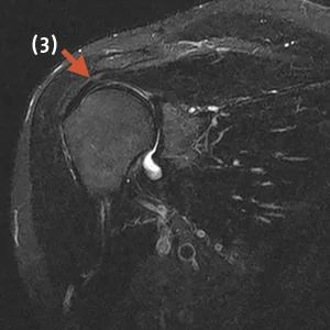
A cuff of fluid is seen around the long biceps tendon (4), most likely indicative of mild tendinitis.
The initial suspicion of rotator cuff rupture was ruled out by magnetic resonance imaging (MRI) as a result of the shoulder examination performed in the Upright MRI, which was fully tolerated by the patient.