Ehlers-Danlos syndromes (EDS) are among the rare diseases, affecting one in about 5000 people. It is a diverse group of congenital connective tissue diseases. The structure of the connective tissue is pathologically altered by a genetic defect.
EDS is a multi-system disease with various involvement of the musculoskeletal system, skin, blood vessels, nerve (pathways), sensory organs, internal organs, etc….
EDS is a hereditary disease, in which mutations are present in the genes responsible for the production of connective tissue. Most of them affect the collagen formation, a group of proteins (proteins), which even form the most common protein in the human body with about 30% share.
Collagens provide tensile and tear strength to the connective tissue found throughout the body and serve as a support, protective and connecting scaffold.
In EDS, genetic defects lead to a disturbed composition of the connective tissue. This becomes too elastic and brittle and can therefore no longer provide a sufficient support function.

Hypermobile Ehlers-Danlos syndrome
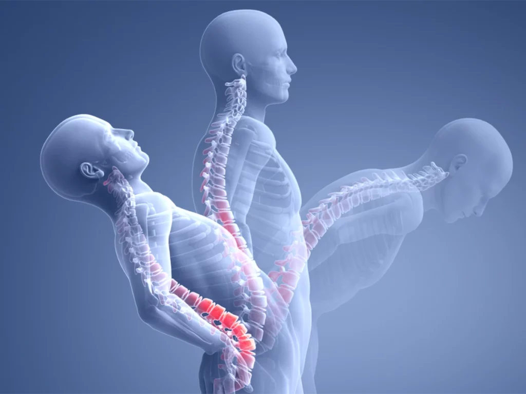
In the special, frequently occurring hypermobile type (hEDS), there is a general overmovement of the musculoskeletal system (bones, joints, tendons, ligaments, cartilage, muscles, fascia) and a reduced physical load-bearing capacity, which leads to considerable problems for many affected persons – up to and including disability.
The symptoms of hEDS are many and vary greatly: changes in the spine (herniated discs, instabilities, blockages, hypermobility, etc.), nerve crushes and (sub)dislocations of joints, frequent soft tissue injuries such as tendon ruptures or muscle tears, inflammation of tendons and bursae, acute and chronic pain, chronic fatigue and exhaustion (CFS, chronic fatigue syndrome), to name a few.
Upright MRI examinations and Ehlers-Danlos syndrome
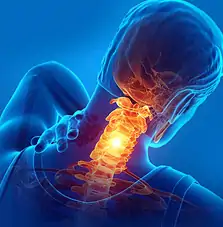
Upright MRI scans of the cervical spine (CS) can be particularly helpful in diagnosing and investigating the symptoms of Ehlers-Danlos syndrome.
An upright MRI scan can provide a much more thorough examination of hEDS patients because it includes a dynamic element that is absent from conventional tunnel MRI scans, in which patients are typically examined while lying down.
Advantages of an examination in functional position in Ehlers-Danlos syndrome (EDS)
The MRI examination of the cervical spine in the Upright MRI is performed in an upright sitting position with the neck in a normal position, then flexed and then extended to obtain a dynamic examination with the unique possible image sequence.
Upright MRI examination of the upper cervicals is one of the system’s clinical specialties and provides an upright examination to allow a complete evaluation of the foramen magnum and cerebellar tonsils (these are structures in the brain located at the base of the cerebellar hemispheres, two halves of the cerebellum (the part of the brain that controls movement and balance).
The two examinations necessary for diagnosis take about 45min each.
The cerebellar tonsils are clearly visible and can be assessed in more detail during an upright examination.
MRI examinations are performed in an open system in different views and different positions:
For example, the head rotated to the right, forward, and to the left, resulting in a rotational dynamic examination. The neck is then also examined in flexion, extension and neutral position. Static measurements are then taken and compared with standardized normal values.
Further measurements are taken in flexion and extension to assess the degree of movement of the person. The ligaments (bands in the upper cervical region) are one of the most important areas examined to determine if they are slack or damaged.
Other advantages of an upright MRI scan of the upper cervical joints and cervical spine include the following
- Fully open upright MRIs are a more comfortable and stress-free alternative to traditional closed tunnel MRIs, offering more comfort and safety for people who suffer from anxiety and/or claustrophobia. Sitting upright is more comfortable for patients, and the open front means patients can talk to a friend or relative during the exam or watch TV for distraction.
- Upright MRI examinations are also suitable for larger / heavier patients who normally do not fit into a conventional tunnel MRI, as they can support a weight of up to 230 kg. Of course, the suitability also depends on the patient’s stature.
The examinations of the upper cervical joints and cervical spine for the diagnosis of hEDS are performed in the practices for upright MRI in Munich, Frankfurt, Hamburg and Hanover: