The 60-year-old patient presented here complains of chronic lumbar syndrome with lumbago, tension, and pseudoradicular lumboischialgia with bilateral radiation to the legs. These complaints occur especially during standing and walking. There is a limitation of walking distance with a feeling of powerlessness in the legs. A conventional MRI examination in the supine position revealed a spinal stenosis due to a prolapse at L3/L4.
Due to the position-dependent discomfort symptoms, kinetic positional magnetic resonance imaging was also performed.
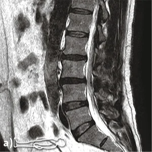

(a) supine image: spinal stenosis in suspected disc herniation with inconspicuous trailing edge alignement in segment L3/L4.
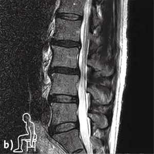
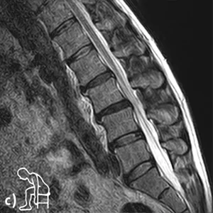
(b + c) Functional images in sitting position: Additionally demonstrable functional anterolisthesis in combination with angular instability in segment L3/L4, but no disc prolapse.
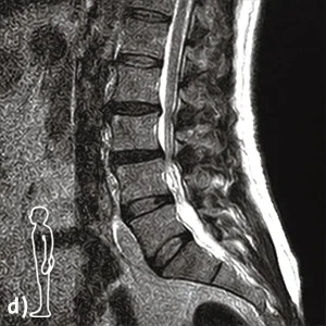
(d + e) Functional images in standing position: Maximally pronounced spinal stenosis with combined lumbar instability with inconspicuous posterior edge alignement in segment L3/L4.
Findings:
Kinetic-positional magnetic resonance imaging under natural weight-bearing showed a dorsal tear of the annulus fibrosus and a circular disc protrusion extending into the neuroforamina with accentuation in reclination posture and regression in inclination posture, in addition to moderate osteochondrosis, in the L2/L3 segment.
In the segment L3/L4 a pseudospondylolisthesis with a ventral displacement from L3 to L4 of about 5 mm was shown in the images under natural weight bearing in neutral and inclination posture. The reclination posture in the standing position and the supine images showed an inconspicuous posterior edge alignment.
In addition, the L3/L4 segment showed angular instability with dorsal
increased hinging in the disc space of about 10 degrees between inclination and reclination posture as well as a function-dependent absolute spinal canal stenosis at the level of the disc, especially accentuated in reclination posture in standing position with an a.p. diameter of 4mm compared to a normal spinal canal width in inclination posture.
There was also a marked narrowing of both neuroforamina, emphasized in reclination.
Diagnosis:
Absolute functionally dependent spinal canal stenosis in L3/L4 in reclination in standing position with pincer-like constriction of the cauda fibers. Combined lumbar instability in segment L3/L4 in pseudospondylolisthesis with functional anterolisthesis in neutral and inclination position in sitting as well as angular instability with dorsally increased hinging in the disc space L3/L4 in inclination position.
Relative function-dependent spinal stenosis in L2/L3.
Upright MRI provided a definitive diagnosis that could not be made by conventional supine MRI alone.