The patient presented here suffered a cervical whiplash injury in a traffic accident about 40 years ago. Since that time she has had migraine-like headaches. For the past 1.5 years, she has had increasing limitations of cervical spine mobility and pain in the craniocervical junction. Since January of this year, she has also had tinitus on the left side and frequent tingling sensations in her left hand.
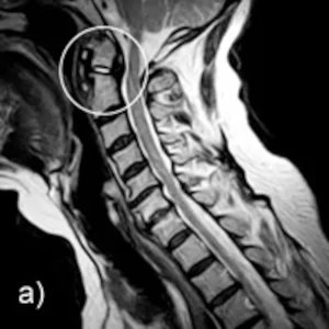
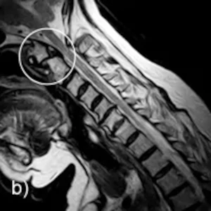
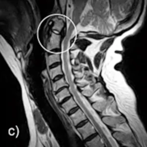
A-C: Sagittal images in the sitting position: Neutral position, inclination, reclination
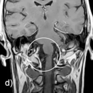
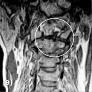
D-E: Coronal image in sitting position, head neutral
Findings: Upright™ MRI of the head reveals abnormalities of the dens axis. Examination of the cervical spine, upright under natural weight bearing, reveals a cleft in the axis at the base of the dens. Functional imaging in Upright™ MRI can demonstrate displacement of the cranial portion of the dens. The sagittal image in the inclination position clearly shows a ventral displacement of the cranial part of the dens. Retrodentally, the sagittal image shows increased soft tissue and the coronal image also shows a marginal bulge at the right atlantoaxial joint. Overall, there is a position-dependent, slight spinal tightness at the level of C1/C2.
The diagnosis: old, unstable dens fracture type II according to Anderson and d’Alonzo with resulting function-dependent, minor spinal canal stenosis….