The Upright MRI procedure enables magnetic resonance imaging diagnostics upright under the natural weight load. This makes it possible to visualize pathologies that cannot be depicted with conventional supine images.
The patient presented here has been complaining of pain in the area of the lumbar spine with radiation into the left lateral upper and lower leg for 6 months. The patient further describes a clear increase of the complaints after prolonged standing, at night, however, there is a clear improvement. For clarification, an MRI of the lumbar spine was performed. The images taken while the patient was lying down did not reveal any clear pathological findings. The vertebral bodies are in a regular position in relation to each other. A herniated disc was not found. This was followed by an examination in the upright MRI.
.
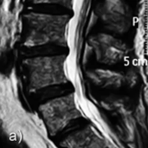
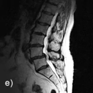
A: Conventional supine imaging in the tunnel system shows no clear indication of the cause of the complaints
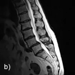
B-C: Sagittal image in sitting position in inclination and reclination
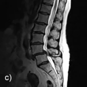
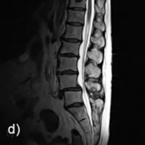
D-E: Sagital image in neutral upright posture in sitting and standing position.
Findings: Upright MRI images were obtained in the sitting, neutral, inclination, and extension positions, with additional sequences in the standing position. The upright images under natural weight bearing show a ventral displacement of L4 compared to L5 in the sense of a degenerative spondylolisthesis (pseudospondylolisthesis) in severe bilateral hypertrophic spondylarthrosis. The spinal canal is highly constricted as a result.
The intervertebral disc L4/L5 shows pathologically increased mobility.