The patient presented here complains of pain lumbar, which can occur in any position, as well as sudden onset pain in the left leg, showing radiation along the L5 root dermatome. There is occasional numbness of the dorsum of the foot.
Upright kinetic-positional MRI was performed for clarification.
In the supine position (Fig. a), there is a slight ventral displacement (4 mm) from L4 to L5. The cause is severe spondylarthrosis with discrete subluxation of the small vertebral joints. Fluid inclusions are found in both small vertebral joints (Fig. b). On the left side, there is a ventrally directed synovial cyst filling the left recessus.
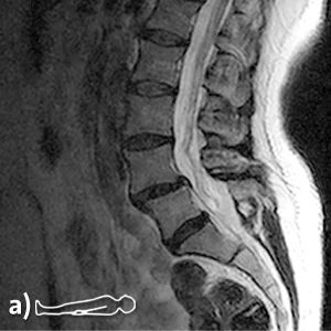
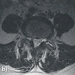
Standard exposures in supine position, a) sagittal T2, b) axial T2
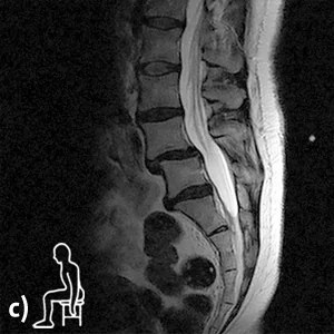
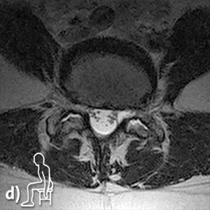
Functional radiographs in sitting position under natural weight load, c) sagittal T2, d) axial T2
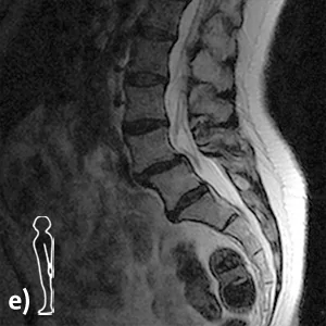
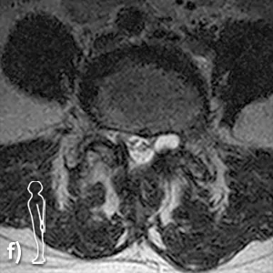
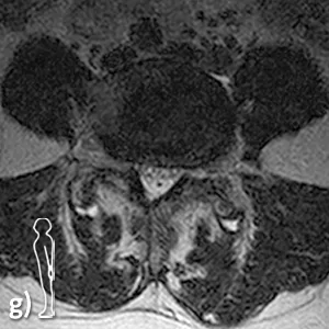
Functional radiographs in standing position under natural weight load, e) sagittal T2, f,g) axial T2
Upright MRI functional examination under the natural weight bearing condition.
In the sitting position, the ventral displacement increases slightly (7 mm), and there is a discrete kyphotic deformity in the segment (Fig. c). The synovial cyst on the left is slightly more prominent; fluid can be detected in the small vertebral joint on both sides.
In the standing position (corresponds to the reclination position), the ventral offset is the same as in the sitting position (7mm) (Fig. e). The joint space is now no longer filled with fluid. On the one hand, this fluid has shifted ventrally into the synovial cyst (Fig. f), and on the other hand, smaller synovial cysts can now be delineated on both sides dorsally of the small vertebral joints (Fig. g).
Diagnosis / Findings:
Low instability (3 mm range of motion) degenerative spondylolisthesis (maximum: 7 mm) in severe spondyloarthritis with discrete subluxation of the small vertebral joints due to severe spondyloarthritis with fluid inclusions.
Weight- and position-dependent synovial cyst in the L4/L5 left recessus caused by spondyloarthritis, communicating with the L4/5 left joint space.
The increase in size of the synovial cyst in the left L4/L5 recess explains the position-dependent L5 root symptoms.
Conventional MRI in the supine position alone cannot depict this.