The 65-year-old female patient presented here has been suffering from complaints in the lumbar spine for many years, which have worsened significantly in the last two years. No side preference, no trauma. Pain radiation into the ventral / ventrolateral thigh on both sides, on the right side significantly stronger than on the left. Increase in discomfort under stress, especially during sports.
A recent MRI examination in the supine position did not provide a clear indication of the cause of the pain symptoms. Subsequently, the patient was examined in the Upright™-MRI, sitting upright under function.
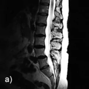
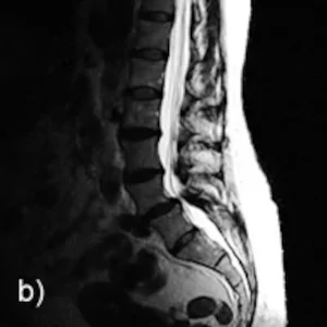
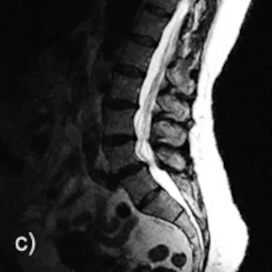
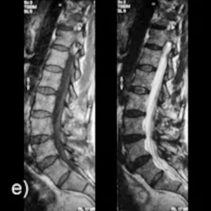
A-C: Sagittal images in the sitting position: Neutral position, inclination, reclination
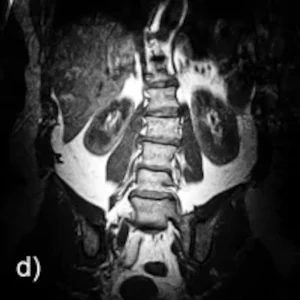
D: Coronary image in sitting position, neutral position
E: Conventional radiograph lying in a tunnel system. Vertebral slippage is not detectable
Diagnosis: Upright MRI examination, upright under natural weight bearing, demonstrated ventral displacement of the L4 vertebral body relative to L5, which was not detectable on conventional supine MRI examination. During the transition to inclination, increasing anterolisthesis is visible at L4/L5 with decrease of the ventrodorsal diameter of the spinal canal at this level. In the upright position, flat disc protrusions are visible at the L2/L3 and L3/L4 levels compared to the supine images. In the coronal sectional plane, a clear left convex lateral malposition of the lumbar spine can be seen.