The 44-year-old female patient presented here complains of complaints in the shoulder region on both sides and a diffuse tingling feeling in the upper extremities on both sides. There was no vertigo and no headache.
Upright magnetic resonance imaging of the cervical spine was performed to clarify the symptoms.
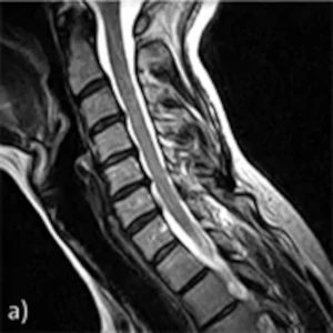
A: Sagittal images in supine position
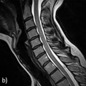
B: Sagittal image in sitting position in neutral posture
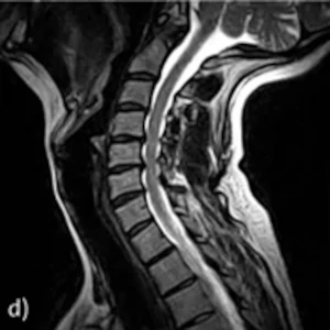
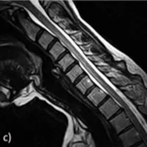
C-D: Sagittal image in sitting position in inclination and reclination
Findings: A standard supine MRI scan (Fig. a) showed shallow dorsal protrusions of the intervertebral discs in the C4/C5 and C5/C6 segments with no evidence of spinal stenosis or nerve root compression at these levels.
In the neutral position in the upright sitting position (Fig. b), there were no significant changes in disc conditions at C4/C5 and C5/C6 compared with a standard MRI examination in the supine position.
In the inclination position (Fig. c), only evidence of very shallow disc protrusions dorsally was possible at C4/C5 and C5/C6. The ventrodorsal diameter of the central portions of the spinal canal at these heights was expanded compared with the images in the upright neutral position.
In the reclination position (Fig. d), however, the spinal canal diameter decreased due to an increase in the size of the disc protrusions.
Upright MRI functional studies in inclination and reclination show position- and motion-dependent polysegmental spinal stenosis from C4/C5 to C6/C7.