A 61-year-old female patient presented with a postoperative persistent lumbar syndrome after long spondylodesis of L2 to L5 with additional cage implantations and recurrent, clearly load- and position-dependent lumboischialgia with right-sided radiation, especially in sitting and standing, with relative absence of symptoms in the supine position.
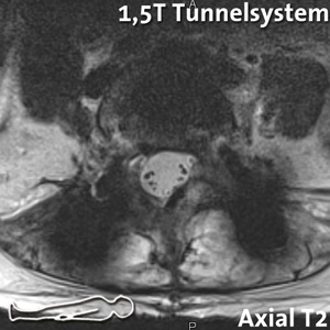
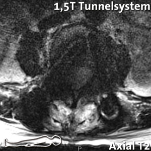
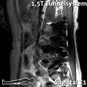
A postoperative supine MRI scan in a 1.5 Tesla tunnel system was not diagnostically useful due to massive metal cancellation artifacts.
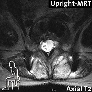
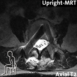
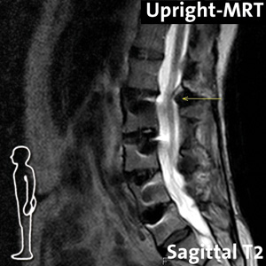
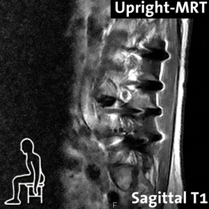
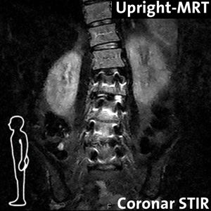
Therefore, a specific presentation for Upright MRI was made in case of clearly load- and position-dependent discomfort symptoms and as a result of the significantly reduced metal artifact formation.

Diagnosis:
The performed kinetic-positional upright MRI examination provides information about the cause of the complaints and shows an atypical medialized position / DD: malposition of the right pedicle screw at the level of L3 with possible irritation of the right L3 nerve root. Otherwise correct position of the osteosynthesis material with clear lumbar scoliosis.
At the level of L4/L5, a paramedian disc prolapse with accentuated adjacent relationship to the right L4 nerve root and consecutive irritation possibility under natural weight bearing in sitting and standing is evident.
Connective degeneration in the segment L1/L2 with formation of posterior instability with demonstrable structural disturbance at the posterior spinal canal boundary in reclination position in standing position and consecutive relative function-dependent spinal canal stenosis.
Due to the extremely low metal artifact sensitivity and the functional images under natural weight bearing in the Upright MRI, a clear diagnosis could be made that explained the patient’s complaint.