The Upright MRI procedure enables magnetic resonance imaging diagnostics upright under the natural weight load. Likewise, complete motion studies are possible with the Upright™-MRI. For example, the spine can be examined under inclination, in reclination, in lateral flexion, or even in rotation, in addition to the neutral, upright position. This makes it possible to visualize pathologies that cannot be depicted with conventional supine images.
The patient presented here never described any problems with the cervical spine until June 2006. Then there had been a sudden onset of fluctuating vertigo that had lasted for about 10 minutes. The patient did not complain of pain in the cervical spine. In January 2007, there had been another attack of vertigo. Since that time also occasional complaint of headache.
A functional examination of the cervical spine was performed with special attention to the cervicooccipital junction on upright MRI.
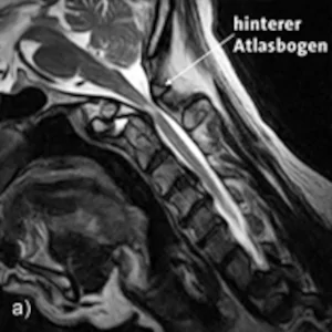
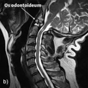
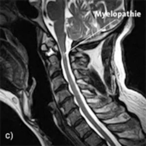
A-C: Sagittal images in the sitting position: Inclination, reclination and neutral position
Findings: Upright™ MRI functional images reveal more severe instability at the cervicooccipital junction with circumscribed pincer-shaped entrapment of the myelon in the inclination position.
A congenital aplasia of the dens is conspicuous, which is only formed as a small bone core in the area of the intended dens tip (Os odontoideum). In the inclination position, there is obviously a circumscribed displacement of the atlas forward with respect to the foramen magnum, which causes the considerable narrowing of the spinal canal at the level of the superior dens. In reclination, the dorsal displacement of the atlas relative to the epistropheus becomes apparent, with marked decompression of the myelon. Circumscribed myelopathy is demonstrable in the region of the superior dens, at the level of the ligamentum transversum.
Incidental findings show disc degeneration on the levels C2/C3 to C5/C6 with shallow disc protrusions, which, however, have no clinical relevance.