The patient presented here underwent surgery in 1995 for a large disc herniation in the L5/S1 segment, which had resulted in paralysis of both legs. Following surgery, the paresis of the legs has completely regressed; however, the patient suffers from persistent back pain.
For the past two years, the pain has increased considerably, radiating into both calves, more so on the right than on the left. The patient finds it particularly difficult to stand and walk, while she is largely symptom-free when lying down.
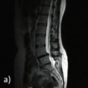
A: Sagittal images in supine position
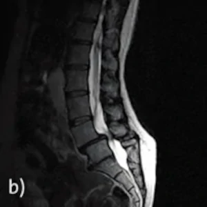
B: Sagittal image in standing position
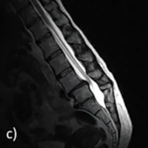
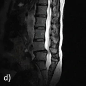
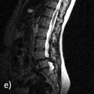
C-E: Sagittal image in sitting position: Inclination, neutral position, reclination
The findings: A standard MRI examination performed in the supine position (Fig. a) showed, like a previous examination in November 2006, a harmonious lordosis of the lumbar spine. Clear median protrusion of the disc L4/L5 into the spinal canal with chondrosis. There is also chondrosis and height reduction of the L5/S1 disc. The disc compartments from L1/L2 to L3/L4 are regular.
The images in standing position (Fig. b) show a significant ventral displacement from L4 to L5 of 9 mm in severe hypertrophic spondylarthrosis. Due to the ventral displacement, a highly severe spinal constriction occurs on this floor.
The functional examination was performed in sitting position in inclination (Fig. c), neutral position (Fig. d) and reclination (Fig. e). In reclination, as in standing, there is an accentuation of spinal tightness with small vertebral joints pressing forward. Beyond that, there is no pathological mobility. The segment L5/S1 shows no mobility in the functional images. Neuroforaminal tightness is not found in any position.
The examination shows a highly load-dependent spinal narrowing at L4/L5 caused by a ventrolisthesis from L4 to L5 in severe hypertrophic spondyloarthrosis.
The findings are to be interpreted as connection instability with segment L5/S1 no longer mobile.
Such a finding cannot be visualized by conventional supine MRI alone.