The 34-year-old patient presented here complains postoperatively of increasing load- and position-dependent pain with radiation into both legs, numbness in the soles of the feet, and tingling paresthesias on the right side, especially in anteflexion. No complaints when lying down. Multiple operations in the area of L5/S1, last hemilaminectomy 5 months ago. No confirmed symptom-free postoperative interval.
A 4-month-old prone MRI scan did not yield a definitive diagnosis. The presentation in the Upright MRI was for clarification of a functional stenosis.
Upright MRI examinations were performed under natural weight bearing in a neutral upright sitting position, as well as in flexion in a sitting position, extension in a standing position, and without weight bearing in a neutral supine position. Four-month-old images from an external supine examination were available for comparison.
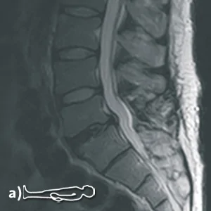
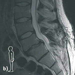
a) Sagittal T2 while lying down, b) Sagittal T2 while standing up.
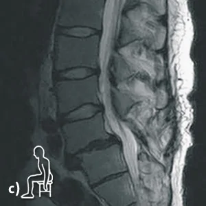
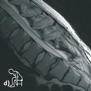
c) Sagittal T2 in sitting position, d) Sagittal T2 in inclination
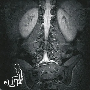
e) Coronary STIR sequence in sitting position with right convex scoliosis in the lower lumbar region with pelvic obliquity.
Diagnosis:
The four-month-old examination available for comparison, as well as the supine upright MRI examination performed on the same day, do not show a clear explanation for the patient’s continued postoperative discomfort symptoms.
Only the kinetic-positional Upright MRI performed provided information about the complete cause of the complaints:
The segment L5/S1 shows a ventrolisthesis from L5 to S1 of 7 mm in anteflexion with a decrease to 5 mm in extension posture in standing position, which can be interpreted as a clearly unstable ventrolisthesis with accompanying angular instability.
Broad-based disc protrusions on all sides with contact to the L5 nerve roots and pelotting of the dural sac at the junction of S1 on both sides. Mild to moderate spinal stenosis. Moderate neuroforaminal stenosis on the right and high-grade neuroforaminal stenosis on the left with further functional increase in anteflexion.
While the spinal as well as the neuroforaminal stenoses are visualized in the supine images, the supine and the standing images do not show the unstable ventrolisthesis from L5 to S1, which is only fully visualized in the anteflexion position with an offset of 7 mm.
Upright MRI functional images provided a definitive diagnosis that could not be made by conventional supine MRI.